Introduction
It is very important to identify the underlying cause of ear symptoms such as tinnitus and hearing loss. If these symptoms present unilaterally, a retocochlear lesion, vascular lesion or inner ear anomaly should be considered in the diagnostic process. Many diagnostic tools have been suggested for use in the evaluation of retrocochlear lesions. However, there is no single novel method to easily evaluate all lesions causing unilateral ear symptoms.
Since Selters first introduced auditory brainstem evoked response (ABR) as a diagnostic method for retrocochlear lesions, the ABR test has been commonly used as a screening test. However, the low sensitivity of the ABR test has lowered its diagnostic value as a screening test, especially in small intracanalicular tumors.1,2)
After the introduction of magnetic resonance image (MRI) technology, MRI testing was applied in the assessment of retrocochlear lesions with high sensitivity and high specificity.3) Nevertheless, the high cost of conventional MRI testing, the use of contrast media and the long image acquisition time might be limitations for using it as a screening tool.
Recently, three-dimensional fast imaging employing steady-state acquisition (3D-FIESTA) images have been introduced. 3D-FIESTA imaging can give much higher spatial resolution with outstanding image contrast between the cranial nerves and cerebrospinal fluid. And it has a shorter image acquisition time than conventional MRI scan and does not need contrast media.4,5)
In this study, we aimed to evaluate the usability of 3D-FIESTA imaging as a screening tool for inner ear lesions with applying 3D-FIESTA image in the diagnostic pathway of the patients with unilateral ear symptoms.
Subjects and Methods
From January 2011 to December 2011, 253 patients who presented with unilateral ear symptoms and underwent 3D-FIESTA imaging as a screening were enrolled in this study. Patients suspected to have Meniere's disease and complained of bilateral ear symptoms were excluded. A thorough medical review of the clinical presentation and audiometric results was performed retrospectively.
All patients underwent 3D-FIESTA imaging as a screening tool with a Philips Achieva (Best, the Netherlands) 3.0 tesla MRI scanner. If an inner ear lesion or a brain lesion was suspected on the 3D-FIESTA image, an additional diagnostic brain MRI scan, a temporal MRI scan, or temporal bone CT scans were performed to elucidate the final diagnosis.
The images from the screening and diagnostic imaging investigations were reviewed to compare their ability to find various cochlear, retrocochlear or vascular lesions.
Patients were divided into three groups according to their chief complaint: tinnitus, unilateral hearing loss, and unilateral sudden idiopathic hearing loss, to compare their clinical presentations and radiological results.
Clinical data and 3D-FIESTA results were analyzed to figure out the relationships between the clinical presentations of acoustic neuroma and tumor size. The Kruskal-Wallis test was applied for the statistical analysis of tumor size using commercially available software (Statistical Package for the Social Sciences, Version 17.0; SPSS Inc., Chicago, IL, USA). A p value of <0.05 was considered as statistically significant.
Results
Among two hundred and fifty-three patients with a unilateral ear symptom, 105 (41.5%) patients were male and 148 (58.5%) were female. The mean age was 50.7±15.7 years.
Tinnitus was the most common symptoms of patients (129, 49.6%), sudden sensory neural hearing loss was the second most common (83, 31.9) and unilateral hearing loss was the next most common (41, 15.7%).
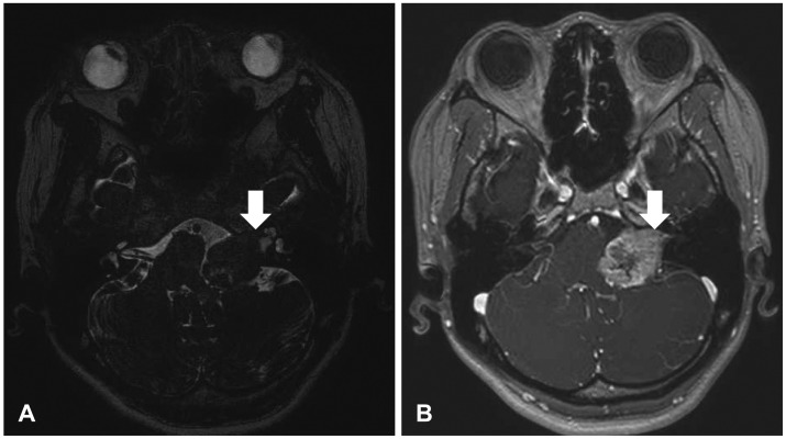
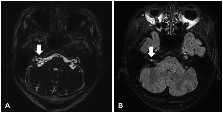
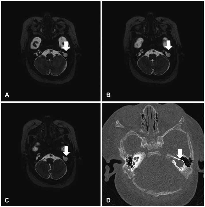
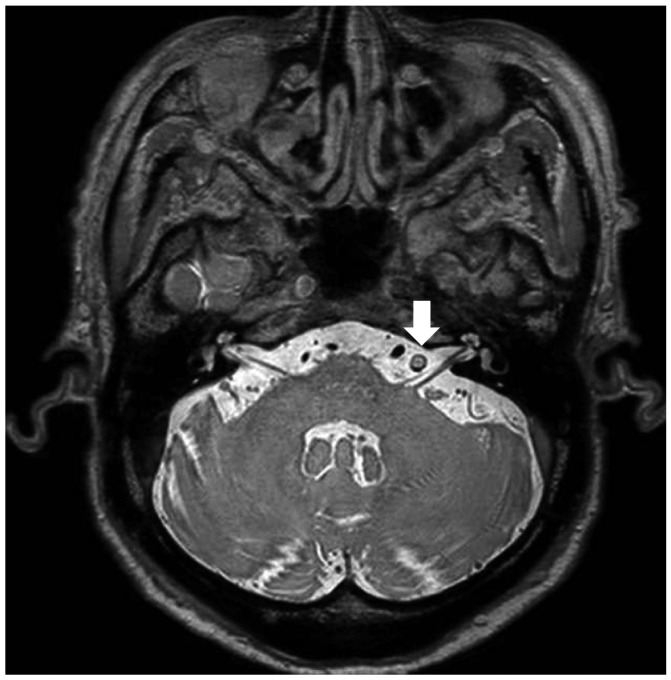
In the 3D-FIESTA imaging results, acoustic neuroma was the most common retrocochlear lesion in patients with unilateral ear symptoms in this study (Fig. 1). Twelve patients were diagnosed with acoustic neuroma (Figs. 2-4), and four patients were confirmed as having enlarged vestibular aqueduct syndrome (Fig. 5). Other lesions such as a congenital anomaly, a vascular lesion, a mass and acute infarction could be detected with 3D-FIESTA imaging (Figs. 6-12).
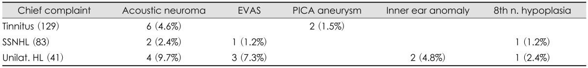
We also tried to figure out the association between clinical symptoms and the final diagnosis. Six patients who initially presented with tinnitus were diagnosed with acoustic neuroma and two patients were diagnosed with posterior inferior cerebellar artery aneurysm. Abnormal findings in 3D-FIESTA images in patients with hearing loss were acoustic neuroma, enlarged vestibular aqueduct syndrome, inner ear anomaly and 8th nerve hypoplasia (Table 1).
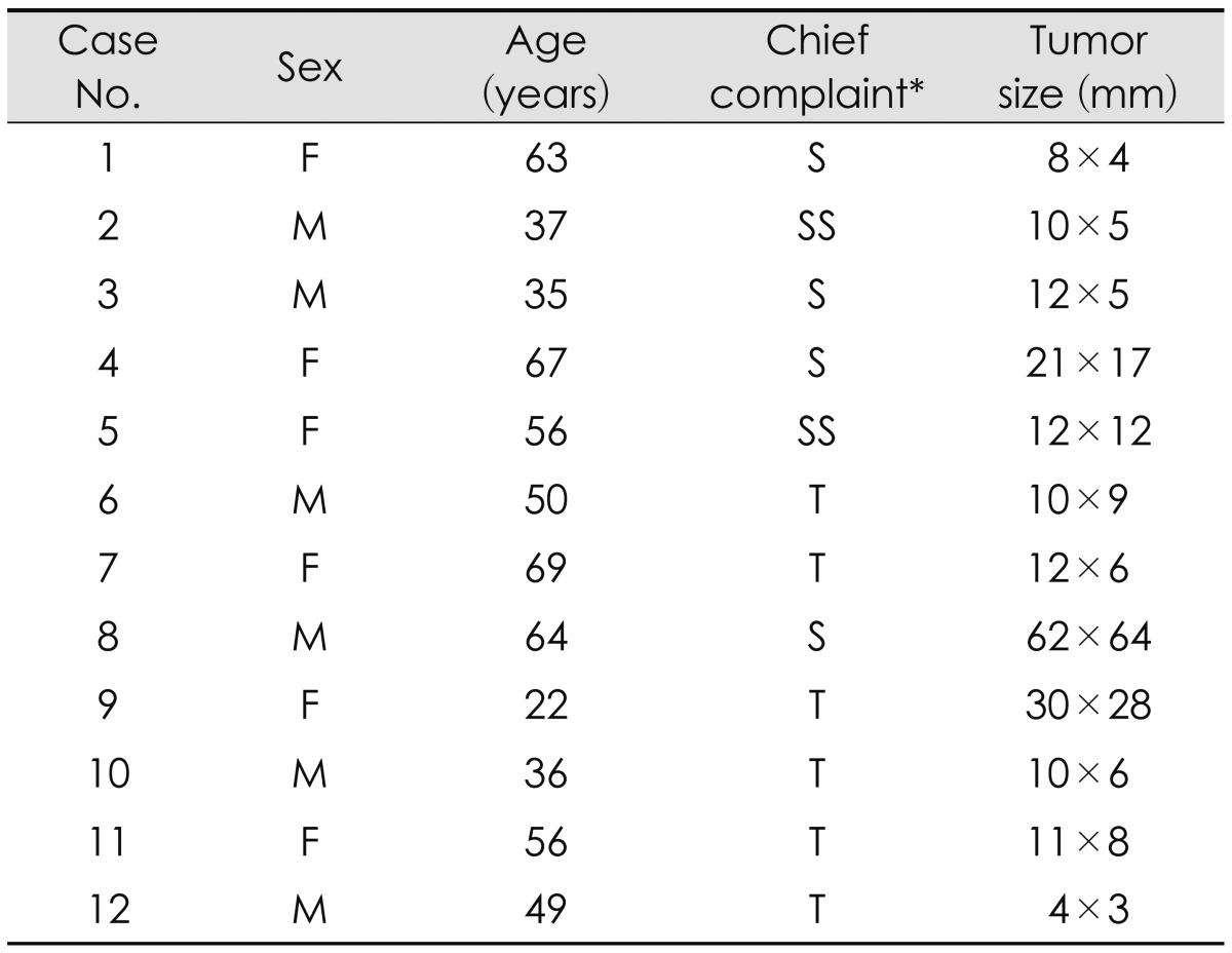
The mean size of acoustic neuromas which were finally diagnosed was 17×14 mm (4×3-62×64 mm), the mean size of tumors in the tinnitus group, unilateral hearing loss group and sudden idiopathic sensorineural hearing loss group was 13×10 mm, 52×23 mm, and 11×9 mm respectively. Table 2 shows the clinical characteristics of patients who were finally diagnosed with acoustic neuroma. The mean tumor size of the unilateral hearing loss group tended to be smaller than that of other groups. However, we could not find any significant correlation between the groups (p=0.582).
Discussion
In patients with unilateral ear symptoms, it is important to approach these patients with a high index of suspicion for cochlear or retrocochlear lesions such as an inner ear anomaly, tumor, vascular or inflammatory lesion.
With conventional audiologic testing, retrocochlear lesions can be suspected by a relatively low speech discrimination score or low score in the short increment sensitivity index test. Retrochoelar lesions were diagnosed more easily after computed tomography (CT) and the ABR test were introduced.6,7) But the temporal bone CT scan is inadequate for screening tool due to the high false or negative positive rate for the diagnosis of retrocochlear lesions especially in the case of acoustic neuroma. ABR test has been used as a favored screening tool for retrocochlear lesions; this test compares the amplitude and latency of waves produced by the auditory pathway. However, Barrs, et al.8) reported that even though ABR demonstrated 97% of the tumors larger than 1.5 cm, 48% of neuromas are 1.5 cm or smaller. Ruckenstein, et al.9) reported that the ABR screening test for retrocochlear pathology had a sensitivity of 63% and a specificity of 64%. These findings imply that ABR probably has a limited role in acoustic neuroma screening.
After the application of MRI for the diagnosis of retrocochlear lesions began in the 1980s, the specificity and sensitivity of MRI was found to be up to 100% and it became to be considered the most accurate diagnostic tool.10,11) But MRI is a very expensive test and it takes 40-60 minutes to get images with contrast. Thus, MRI has limitations as a screening test due to the low cost and time effectiveness.
With the development of the image acquisition technique with MRI, new protocols for 3D-FIESTA sequences were introduced which provided much higher spatial resolution and clearer depiction of small structures such as cranial nerves especially within the cisternal spaces. The 3D-FIESTA uses an ultrafast pulse sequence that produces 0.3 mm thin section high resolution images with outstanding image contrast between the cerebrospinal fluid, vessels and cranial nerves. It usually takes 5 to 7 minutes to get a proper image and does not need contrast media. The cost of 3D-FIESTA is just 1/5-1/6 that of conventional MRI. So it could be a very effective test protocol for the screening of retrocochlear lesions in patients with unilateral ear symptoms.
In this study, variable lesions such as small tumors less than 5 mm, inner ear anomalies and vascular malformations could be identified using 3D-FIESTA imaging. And there is one report which showed that with 3D-FIESTA, there was decreased vestibular signal intensity on the affected side in patients with vestibular schwannoma, but that this was not present in those with cerebellopontine angle meningiomas or in normal subjects.12)
Recently, MRI with high-dose contrast media has been introduced for detecting cochlear endolymphatic hydrops.13) 3D-FIESTA images have a limitation in detecting inner ear lesions; however, 3D-FIESTA images can detect inner ear anomalies so it may be sufficient to use as a screening tool for retrocochlear lesions.
Acoustic neuroma, one of the most common retrocochlear lesions, is a benign tumor originating from the vestibular nerve sheath in the internal auditory canal. In the early stage of acoustic neuromas, symptoms can be nonspecific but as the tumor grows, progressive unilateral hearing loss and tinnitus are typical symptoms. It is well known that about 10 to 20% of acoustic neuroma patients can be present with sudden sensorineural hearing loss, and on the other hand only 1 to 3% of sudden seonsorineural hearing loss patients have acoustic neuroma.14-16)
In this study, 9.7% of unilateral hearing loss patients, 4.6% of unilateral tinnitus and 2.4% of sudden sensorineural hearing loss patients were diagnosed with acoustic neuroma. The mean size of acoustic neuroma varied from a tiny tumor smaller than 5 mm to a large tumor up to 64 mm. The mean size of the tumor in the sudden hearing loss patients group was larger than the other two groups, but there was no statistically significant difference between the groups. In addition to acoustic neuroma, many retrocochlear lesions, intracranial masses and acute infarctions can be diagnosed with screening using 3D-FIESTA imaging.
With these findings, we suggest that 3D-FIESTA imaging could be a reliable screening test for patients who complain of unilateral otologic symptoms. But considering that this study was a simple retrospective observational study, further comparative study investigating the sensitivity and specificity of 3D-FIESTA imaging will be required.