Warning: mkdir(): Permission denied in /home/virtual/lib/view_data.php on line 81 Warning: fopen(/home/virtual/audiology/journal/upload/ip_log/ip_log_2024-04.txt): failed to open stream: No such file or directory in /home/virtual/lib/view_data.php on line 83 Warning: fwrite() expects parameter 1 to be resource, boolean given in /home/virtual/lib/view_data.php on line 84 Osteoma of the Promontory Mimicking a Congenital Cholesteatoma
Korean J Audiol Search

CLOSE


J Audiol Otol > Volume 18(1); 2014 > Article
Park, Eom, Cho, and Jang: Osteoma of the Promontory Mimicking a Congenital Cholesteatoma

Abstract

Osteoma of the temporal bone most commonly occurs in the external ear. Osteomas in the middle ear are not common, and only 25 relevant cases (18 papers) have been reported in the English literature. With only 5 cases reported to date, osteoma of the promontory in the middle ear is rare. This study reports a case of asymptomatic osteoma of the promontory in the middle ear mimicking a congenital cholesteatoma in a 4-year-old girl.

Introduction

Osteoma of the temporal bone is benign pedunculated tumors of the lamellar bone that are most commonly found in the external auditory canal. Osteomas in the middle ear are not common, and since their first mention,1) only 25 relevant cases (18 papers) have been reported in the literature. In previously reported cases, osteomas in the middle ear were combined with cholesteatoma or ossicular osteoma. However, osteoma of the promontory of the middle ear is even rarer, with only 5 cases reported to date.2,3,4,5,6) This study reports a case of asymptomatic osteoma of the promontory in the middle ear mimicking a congenital cholesteatoma in a 4-year-old girl.

Case Report

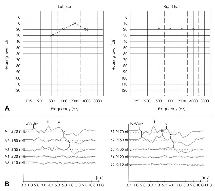
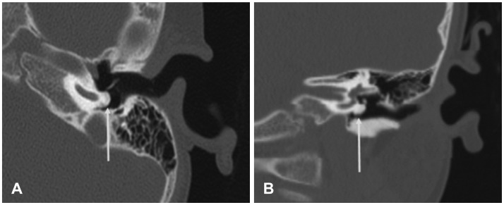
The patient whose case is presented here was a 4-year-old girl without a history of hearing loss and otorrhea. She denied dizziness and tinnitus. Having been diagnosed with congenital cholesteatoma at a local clinic, she was transferred to our department. Otoendoscopic examination revealed a white mass behind the left tympanic membrane mimicking a congenital cholesteatoma (Fig. 1). A MASTER (Bio-logic, San Carlos, CA, USA) system was used for auditory steady-state response, auditory brainstem response measurement. It revealed normal range of hearing (Fig. 2). Temporal bone high resolution computerized tomography was conducted with Discovery CT 750 (GE, New York, NY, USA). Slice thickness is 1.25 mm and it disclosed the presence of bony mass covering the left cochlear promontory at the level of the basal turn (Fig. 3). In radiological examination, this mass was identified as an osteoma. The bony mass did not involve the ossicles, oval and round windows, or obstruction of the Eustachian tube orifice. No further anomalies were observed. This asymptomatic mass was not removed. Periodical follow-up with temporal bone high resolution computed tomography was needed.

Discussion

Osteoma of the temporal bone most commonly occurs in the external ear. On rare occasions, it may arise from other locations within the temporal bone, such as the ossicle (malleus, incus), lateral semicircular canal7) and the petrous ridge.8)
Osteomas on ossicle may cause a conductive hearing loss by impinging on the ossicular chain. However there is no related sensorineural hearing loss.9,10,11,12)
The etiology of the osteoma of the promontory remains unknown. Most of the cases of middle-ear osteomas were asymptomatic and were diagnosed coincidentally. In the present case, the osteoma was asymptomatic. Hearing tests revealed the normal range. If the patient has a conductive hearing loss, surgical intervention can be performed using exploratory tympanotomy. Care should be taken not to avoid damage to the ossicular chain or iatrogenic labyrinthine fistula. In Cremers,2) the promontory osteoma was removed using a chisel, but the traumatic labyrinthine fistula was followed. If the mass is asymptomatic or when it does not involve the ossicular chain, surgical removal is not necessary. Periodical follow-up with HRCT and audiologic examination is needed. In case of progressive growth of the osteoma with an increasing hearing loss, ossicular chain involvement, obstruction of Eustachian tube, or round window obstruction, surgery should be considered.

References

1. Thomas R. Familial osteoma of the middle ear. J Laryngol Otol 1964;78:805–807. PMID: 14205962.
crossref pmid
2. Cremers CW. Osteoma of the middle ear. J Laryngol Otol 1985;99:383–386. PMID: 4009038.
crossref pmid
3. Glasscock ME 3rd, McKennan KX, Levine SC. Osteoma of the middle ear: a case report. Otolaryngol Head Neck Surg 1987;97:64–65. PMID: 3112686.
crossref pmid
4. Silver FM, Orobello PW Jr, Mangal A, Pensak ML. Asymptomatic osteomas of the middle ear. Am J Otol 1993;14:189–190. PMID: 8503496.
pmid
5. Ramírez-Camacho R, Pinilla M, Garcia-Berrocal JR. Osteoma of the promontory: a case report. ORL J Otorhinolaryngol Relat Spec 1997;59:64–65. PMID: 9104753.
crossref pmid
6. Unal OF, Tosun F, Yetişer S, Dündar A. Osteoma of the middle ear. Int J Pediatr Otorhinolaryngol 2000;52:193–195. PMID: 10767470.
crossref pmid
7. Ben-Yaakov A, Wohlgelernter J, Gross M. Osteoma of the lateral semicircular canal. Acta Otolaryngol 2006;126:1005–1007. PMID: 16864503.
crossref pmid
8. Beale DJ, Phelps PD. Osteomas of the temporal bone: a report of three cases. Clin Radiol 1987;38:67–69. PMID: 3816069.
crossref pmid
9. Shimizu T, Okamoto K, Majima Y. Osteoma of the malleus: a case report and literature review. Am J Otolaryngol 2003;24:239–241. PMID: 12884215.
crossref pmid
10. Milroy CM, Phelps PD, Michaels L, Grant H. Osteoma of the incus. J Otolaryngol 1989;18:226–228. PMID: 2769836.
pmid
11. Yamasoba T, Harada T, Okuno T, Nomura Y. Osteoma of the middle ear. Report of a case. Arch Otolaryngol Head Neck Surg 1990;116:1214–1216. PMID: 2206511.
crossref pmid
12. Ramdoo K, Dale OT, Herdman RC. Malleo-incudal osteoma: an unexpected finding during surgery for presumed otosclerosis. J Laryngol Otol 2011;125:968–969. PMID: 21745432.
crossref pmid
Fig. 1
Otoendoscopic view of left tympanic membrane. White mass behind the tympanic membrane is seen at inferior site.
kja-18-38-g001.jpg
Fig. 2
ASSR & ABR. A: Auditory steady-state responses reveal the normal range in both sides. B: Auditory brainstem responses reveal the normal range 30 dB in both side. ASSR: auditory steady-state response, ABR: auditory brainstem response.
kja-18-38-g002.jpg
Fig. 3
Temporal bone CT shows the mass susceptible to osteoma arising from the promontory. A: Axial view: arrow indicates the mass susceptible to osteoma of the promontory. B: Coronal view: arrow indicates the mass susceptible to osteoma of the promontory.
kja-18-38-g003.jpg


ABOUT
ARTICLES

Browse all articles >

ISSUES
TOPICS

Browse all articles >

AUTHOR INFORMATION
Editorial Office
The Catholic University of Korea, Institute of Biomedical Industry, 4017
222, Banpo-daero, Seocho-gu, Seoul, Republic of Korea
Tel: +82-2-3784-8551    Fax: +82-0505-115-8551    E-mail: jao@smileml.com                

Copyright © 2024 by The Korean Audiological Society and Korean Otological Society. All rights reserved.

Developed in M2PI

Close layer
prev next